MITI Minimum Information guidelines for highly multiplexed tissue images
Abstract
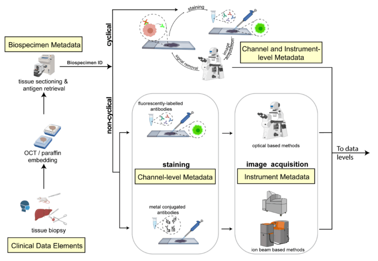
The imminent release of atlases combining highly multiplexed tissue imaging with single cell sequencing and other omics data from human tissues and tumors creates an urgent need for data and metadata standards compliant with emerging and traditional approaches to histology. We describe the development of a Minimum Information about highly multiplexed Tissue Imaging (MITI) standard that draws on best practices from genomics and microscopy of cultured cells and model organisms.
Citation
D Schapiro, C Yapp, A Sokolov, SM Reynolds, YA Chen, D Sudar, Y Xie, J Muhlich, R Arias-Camison, S Arena, AJ Taylor, M Nikolov, M Tyler, JR Lin, EA Burlingame, Human Tumor Atlas Network, YH Chang, SL Farhi, V Thorsson, N Venkatamohan, JL Drewes, D Pe’er, DA Gutman, MD Herrmann, N Gehlenborg, P Bankhead, JT Roland, JM Herndon, MP Snyder, M Angelo, G Nolan, JR Swedlow, N Schultz, DT Merrick, SA Mazzili, E Cerami, SJ Rodig, S Santagata, PK Sorger. “MITI Minimum Information guidelines for highly multiplexed tissue images”, Nature Methods 19(3):262-267 (2021). doi:10.1038/s41592-022-01415-4